FAQ

  • Fibre
  • Ulcerative Colitis
  • Stoma
  • Rectocele
  • Rectal Cancer
  • Pruritus Ani
  • Polyps of the Colon and Rectum
  • Pilonidal Disease
  • Pelvic Floor Dysfunction
  • Irritable Bowel Syndrome
  • Crohn's Disease
  • Colonoscopy
  • Colon Cancer
  • Anal Warts
  • Anal Pain
  • Anal Abscess and Fistula
  • Anal Cancer
  • Haemorrhoids
  • Diverticulosis
  • Rectal Prolapse

Fibre

What is Fibre?

Fibre is an important part of a healthy balanced diet. It can help prevent heart disease, diabetes, weight gain and some cancers, and can also improve digestive health. You should aim for at least 30g a day.

Fibre is only found in foods that come from plants. Foods such as meat, fish and dairy products don't contain any fibre.

There are two different types of fibre – soluble and insoluble. Each type of fibre helps your body in different ways, so a normal healthy diet should include both types. Eating wholegrain cereals and plenty of fruit and vegetables helps to ensure both adults and children are eating enough fibre.

However, if you have a digestive disorder such as irritable bowel syndrome (IBS), you may need to modify the type and amount of fibre in your diet in accordance with your symptoms. Your GP or a dietitian can advise you further about this.

Soluble fibre

Soluble fibre dissolves in the water in your digestive system. It may help to reduce the amount of cholesterol in your blood. If you have constipation, gradually increasing sources of soluble fibre – such as fruit and vegetables, oats and golden linseeds – can help soften your stools and make them easier to pass.

Foods that contain soluble fibre include:

  • oats, barley and rye
  • fruit, such as bananas and apples
  • root vegetables, such as carrots and potatoes
  • golden linseeds

Insoluble fibre

Insoluble fibre doesn't dissolve in water. It passes through your gut without being broken down and helps other foods move through your digestive system more easily. Insoluble fibre keeps your bowels healthy and helps prevent digestive problems. If you have diarrhoea, you should limit the amount of insoluble fibre in your diet.

Good sources of insoluble fibre include:

  • wholemeal bread
  • bran
  • cereals
  • nuts and seeds (except golden linseeds)

Eating foods high in fibre will help you feel fuller for longer. This may help if you are trying to lose weight.

If you need to increase your fibre intake, it's important that you do so gradually. A sudden increase may make you produce more wind (flatulence), leave you feeling bloated, and cause stomach cramps.

It's also important to make sure you drink plenty of fluid. You should drink approximately 1.2 litres (six to eight glasses) of fluid a day, or more while exercising or when it's hot.

Ulcerative Colitis

surgeon mallick

UC can effect the entire colon and rectum

What is ulcerative colitis?

Ulcerative colitis (UC) is an inflammatory disease potentially affecting the entirelarge bowel (colon and rectum). The inflammation is confined to the innermost layer of the intestinal wall (mucosa). UC can go into remission and recur. Medical management is typically the first option for treatment. If surgery is needed for UC, it is usually curative.

What are the symptoms of ulcerative colitis?

The most common symptoms of UC include abdominal cramping, pain, diarrhoea, bleeding with bowel movements, fever, fatigue, and weight loss.

Who is at risk for ulcerative colitis?

UC can appear at any age, but most patients develop symptoms in their 40s, while a smaller proportion can appear later in life (60-70 years of age). Men and women are affected equally. A family history of UC slightly increases the risk of developing it.

What causes UC?

The exact cause of UC is unknown. Current research focuses on abnormalities in the body's immune system and on bacterial infection.

How are patients evaluated?

Initial work up includes a thorough medical history and physical examination followed by laboratory testing including stool tests, completecolonoscopy of the rectum, colon and terminal ileum. This evaluation is important in determining the extent and severity of the disease and guides management. It can be difficult to differentiate ulcerative colitis from a similar disorder, Crohn's disease, when the latter involves only the colon and rectum.

What is the medical treatment for UC?

Medical treatment is always the first choice unless emergency surgery is required. Several treatment plans are available for initial therapy, for maintenance therapy, and to improve the patient's quality of life. The most commonly used initial therapy is corticosteroids combined with anti-inflammatory agents. They can be eithertaken by mouth or as a rectal suppository, depending on the extent of the disease.

surgeon mallick

A (left side): Complete removal of the colon and rectum | B (right side): Creation of an end Ileostomy

What are the available surgical treatments?

Surgery can either be scheduled immediately or at a later date. Emergency surgery is needed for a perforated bowel (hole in the bowel), severe bleeding, or a serious infection (toxic colitis). These conditions potentially threaten the patient's life. Elective surgery is usually for UC that is not responding to medical management or when a cancerous or precancerous change is found during colonoscopy.

Because ulcerative colitis involves only the colon and rectum, complete removal of these with creation of an ileostomy is curative for the disease and is one of the treatment options.

surgeon mallick

Ileoanal Pouch

In emergency surgery for UC, the large bowel (colon) is removed, temporarily leaving the rectum and anus, and the end of the small bowel (ileum) is brought out through the abdominal wall to the skin level to create an ileostomy through which the fecal stream is diverted. Depending upon the particular circumstances, this procedure can be performed in a traditional "open" procedure or in a minimally invasive (laparoscopic) fashion.

surgeon mallick

Complete removal of the colon with creation of end eleostomy. Rectum still in place.

After the patient recovers, a second procedure can be performed to remove the remaining diseased rectum and create a new rectum (ileal pouch) using the small bowel. The new rectum is connected to the anal opening and a loop ileostomy created to protect the area until it has healed.

surgeon mallick

Creation of I-pouch after removal of the rectum with creation of a diverting loop ileostomy.

When healing is complete, a third procedure is done to close the ileostomy. This is the three-stage procedure for UC and ultimately results in patients being able to live without a stoma.

In addition to the risks associated with any surgery and those associated with pelvic surgery, surgery for UC carries a risk that a leak will develop at the surgical connections.

The surgery can be done via either a traditional open or a minimally invasive (laparoscopic) procedure. Your surgeon will help choose your procedure based on which is safest and most appropriate for you. Emergency surgery is most often an open procedure owing to the urgency of the situation.

What follow-up is needed after surgery?

Patients need continuing follow-up appointments with their doctor to evaluate the function and health of the pouch over time.

Stoma

What is a stoma?

Stoma is a surgically created opening between an internal organ and the body surface. Stoma may often be created as a part of intestinal surgery when there must be a new way for waste 
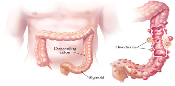
products to leave the body. Cancer, trauma, inflammatory bowel disease (1BD), bowel obstruction, infection, faecalincontinence (inability to control bowel movements) and diverticulitis (inflammation of tiny pockets that commonly form in the colon wall) are all possible reasons for Stoma creation.

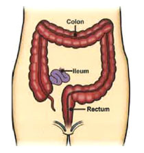
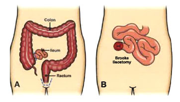
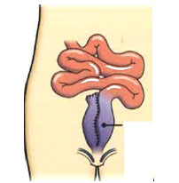
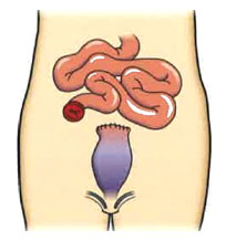
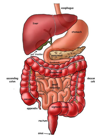
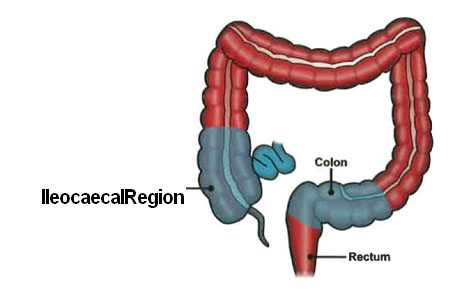
The most common types of stomas include: "ileostomy" (which connects the ileum, the last part of the small intestine, to the skin of the abdominal wall) and "colostomy" (which connects a part of the colon, or large intestine, to the skin of the abdominal wall).

A Stoma may be temporary or permanent. A temporary stoma can usually be reversed. It is created to temporarily keep stool material away from a downstream area (closer to the anus), such as a healing surgery site, an area of inflammation, or a blockage associated with disease or scar. A permanent stoma may be needed when disease, or its treatment, impairs normal intestinal function, or when the muscles that control elimination must be removed or no longer work properly.

How will I control my bowel movements?

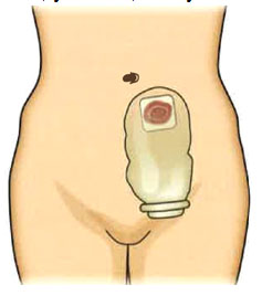
Once a stoma is created, your bowel movements will naturally empty through the stoma. A pouch, called a stoma appliance, is typically worn on the outside of your body around the opening. The appliance isdesigned to catch and hold the emptied stool. A stoma therapist nurse who specializes in stoma care and your colorectal surgeon will teach you how to attach the appliance. The pouch (bag) is made of plastic and is held to the body with an adhesive, which, in turn, protects the skin from moisture. The pouch is disposable and is emptied or changed as needed. The system is quite secure; "accidents" are not common, and the pouches are odour-free.The frequency and quantity of your bowel movements will vary, depending on the type of ostomy you have, your diet, and your bowel habits prior to surgery.

surgeon mallick

A stoma connects either the small or the large intestine to the surface of the body.

Will my diet be limited?

You may be instructed to modify your eating habits in order to control the frequency and consistency of your bowel movements, depending on the type of your stoma. Chew your food well and drink plenty of fluids. You may be asked to avoid certain high roughage foods. Over time, most patients can introduce foods back into your diet a little at a time and monitor the effect of each food on the stoma function. Most patients will eventually have no limitations. Your stoma nurse and colorectal surgeon will give you more details.

Will other people know that I have a stoma?

Not unless you tell them. An stoma is easily hidden by your usual clothing. You probably have met people with a stoma and not realized it!

Where will the stoma be located?

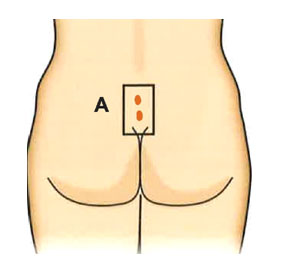
A stoma is best placed on a flat portion of the abdominal wall that can be easily seen and reached by you. Before surgery, it is best for your stoma nurse to mark an appropriate place on your abdominal wall. A colostomy is usually placed to the left of your belly button and an ileostomy to the right.

Will my physical activities be limited?

All your activities, including active sports, may be resumed once healing from surgery is complete. Public figures, footballers, prominent entertainers, and even professional athletes have stoma that do not significantly limit their activities.

Will a stoma affect my sex life?

Most patients with stoma resume their usual sexual activity. Some patients worry about how their sexual partner will think of them because of their stoma appliance. This perceived change in body image can be overcome by a strong relationship, time and patience.

What are the complications of an stoma?

Complications from a stoma can occur. When you first start working with your stoma, it may take some time for you tolearn how the stoma works and how the appliance fits and empties. During this time, there may be occasions when "accidents" happen. Once you have adjusted to the appliance, the most common complications related to the stoma are minor, like local skin irritation, and can be easily remedied.

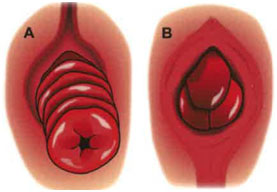
Significant changes in body shape, such asweight loss or gain, can affect the function of a stoma. Other problems that occur over time may include hernia (weakening of the abdominal wall around the stoma) or prolapse (a protrusion of the bowel), that occasionally require surgery if they cause significant symptoms.

Living with a stoma will require some adjustments and learning, but an active and fulfilling life is expected. Your stoma nurse and colorectal surgeon will provide you with skills and support to help you live withyour stoma.

Rectocele

What is a Rectocele?

A rectocele is a bulging of the front wall of the rectum into the back wall of the vagina. The rectum is the bottom section of your colon. This is avery common problem that often does not produce symptoms. Other pelvic organs can bulge into the vagina, including the bladder (cystocele) and the small intestines (enterocele), producing similar problems.

Causes

Rectoceles are usually caused by thinning of the rectovaginal septum (the tissue between the rectum and vagina) and weakening of the pelvic floor muscles. There are many things that can lead to weakening of the pelvic floor, including:

  • Vaginal deliveries.
  • Trauma from vaginal delivery (e.g., the use of forceps or vacuum during delivery, tearing or episiotomy, which is a surgical cut in the muscular area between the vagina and the anus made just before delivery).
  • History of constipation.
  • Chronic straining with bowel movements.
  • Gynaecological (e.g., hysterectomy) or rectal surgeries.

Symptoms

Most people with a small rectocele do not have symptoms. When the rectocele is large, there is usually a noticeable bulge into the vagina.

Rectal Symptoms

  • Difficulty having a complete bowel movement.
  • Stool getting stuck in the bulge of the rectum.
  • The need to press against the vagina and/ or space between the rectum and the vagina to have a bowel movement.
  • Straining with bowel movements.
  • The urge to have multiple bowel movements throughout the day.
  • Constipation.
  • Rectal pain.

Vaginal Symptoms

  • Pain with sexual intercourse (dyspareunia).
  • Vaginal bleeding.
  • A sense of fullness in the vagina.
surgeon mallick

Female Anatomy with Rectocele

Diagnosis

Colorectal surgeons as well as gynaecologists are trained in the diagnosis and treatment of this condition. A rectocele is often found during a routine physical examination. However, other tests maybe needed to help evaluate its severity or possible connection to symptoms. The following test may be ordered to confirm the diagnosis.

Proctogram: A special X-ray test that shows the rectum and anal canal as they change during defaecation. This study is very specific and can pinpoint the size of the rectocele and the degree to which the rectum is emptied.

Treatment Methods

Rectocele treatment is needed only when they cause symptoms that interferewith daily living. Before any treatment, your colorectal surgeon will assess whether all of your symptoms are related solely tothe rectocele. There are nonsurgical and surgical treatment options for rectoceles. Most symptoms associated with arectocele can be resolved with nonsurgical treatment; however, this depends on the severity of symptoms.

Non-Surgical Treatment

The goal is to have good daily bowel habits and softer stools. Avoiding constipation and straining with bowel movements will reduce the risk of a bulge associated with a rectocele.

Preventive and Medical Tips

  • Eating a high-fibre diet and taking over-the-counter fiber supplements (25-35 grams of fiber/day).
  • Drinking more water (typically 6-8 glasses daily).
  • Avoiding excessive straining with bowel movements.
  • Applying pressure to the back of the vagina during bowel movements.
  • Pelvic floor exercises.
  • Biofeedback, a special form of pelvic floor physiotherapy aimed at improving rectal sensation and pelvic floor muscle contraction.
  • Stool softeners.

Surgical Treatment

The surgical management of rectoceles should only be considered when nonsurgical methods have not resolved or improved symptoms and the condition interferes with daily living. This can be done through abdominal, rectal or vaginal surgery. The choice of procedure depends on the size of the rectocele and its symptoms. The goal of surgery is:

  • To remove the extra tissue caused by the rectocele.
  • To strengthen the wall between the rectum and vagina with surrounding tissue or use of a mesh patch.

Post-Surgery

The success rate of surgery depends on the specific symptoms and their duration. Risks of surgical correction include bleeding, infection and pain during intercourse (dyspareunia). There is also the chance of the rectocele recurring or worsening.

Rectal Cancer

surgeon mallick

Rectal Cancer

What is Rectal Cancer?

Rectal cancer arises from the lining of the rectum. The rectum is the bottom 6 inches of your colon (large intestine). Like colon cancer, rectal cancer is highly curable, if detected in early stages. In comparison, colorectal cancer is referring to any cancer that may occur in the colon or rectum.

Risk Factors

The exact cause of rectal cancer is unknown. The following factors can increase one's risk of rectal cancer.

  • Age: More than 90% of people are diagnosed with colon cancer after age 60
  • Family history of colorectal cancer (especially parents or siblings)
  • Personal history of Crohn's Disease or ulcerative colitis for 8 years or longer
  • Colorectal polyps
  • Personal history of breast, uterine or ovarian cancer

Prevention

Rectal cancer is preventable. Nearly all cases of rectal cancer develop from polyps. These polyps are benign growths on the inner lining of the rectum. Detection and removal of polyps through colonoscopy reduces the risk of rectal cancer. Rectal cancer screening recommendations are based on medical and family history. Screening typically starts at age 60 in patients with average risk. Those at higher risk are usually advised to receive their first screening at a younger age.

While it is not definitive, there is some evidence that diet may play a significant role in preventing colorectal cancer. A diet high in fibres (whole grains, fruits, vegetables and nuts) and low in fat may help prevent colorectal cancer.

Symptoms

Rectal cancer often causes no symptoms and is detected during routine screening. It is important to note that other common health problems can cause some of the same symptoms. For example, haemorrhoids are a common cause of rectal bleeding but do not cause rectal cancer.

Rectal cancer symptoms include:

  • A change in bowel habits (e.g., constipation or diarrhea)
  • Narrow shaped stools
  • Bright red or very dark blood in the stool
  • Ongoing pelvic or lower abdominal pain (e.g., flatulence, bloating or pain)
  • Unexplained weight loss
  • Nausea or vomiting
  • Feeling tired all the time

Abdominal pain and weight loss are typically late symptoms, indicating possible extensive disease. Anyone who experiences any of the above symptoms should see a doctor as soon as possible.

Diagnosis

  • Medical history
  • Physical examination
  • Blood tests
  • Digital rectal exam (DRE): Insertion of a gloved, lubricated finger into the rectum to check for abnormalities
  • Proctoscopy: Examination of the anal cavity and rectum using a narrow instrument called a proctoscope
  • Colonoscopy: Examination of the entire colon with a long, flexible instrument called a colonoscope
  • Biopsy: Taking samples of tissues so they can be viewed under a microscope to check for signs of cancer

Rectal Cancer Staging Tests

Distant Staging

  • Computed Tomography (CT) scan:A highly sensitive x-ray test that allow doctors to see "inside" the body and look at all of the organs. This test can help detect the presence of cancer that has spread outside the rectum.
  • CEA assay:Carcinoembryonic antigen is a substance in the blood that may be elevated when cancer is present. Although not completely conclusive on its own, this test is used to help monitor patients after their cancer has been treated.

Local Staging

  • MRI:An imaging test that uses a magnetic field and pulses of radio wave energy to create pictures of organs and structures inside the body. This helps determine if the tumor has spread through the wall of the rectum and invaded nearby structures.

Surgical Treatment

Surgery to remove rectal cancer is almost always required for a complete cure. Depending on the location and stage, this may be performed through the anus (opening of the rectum) or through theabdomen. The tumour and lymph nodes are removed, along with a portion of normal rectum on either side of the tumor. A colostomy is a surgically created openingthat connects a part of the colon to the skin of the abdominal wall. This procedure is typically only done in a very small number of rectal cancer patients.

Keyhole surgery may be used based on the individual case. Your colorectal surgeon will discuss this with you prior to surgery and decide on the best approach.

Medical Treatment

Chemotherapy or radiation therapy may be offered either before or after surgery, depending on the stage of the cancer.

Prognostic (Outcome) Factors

Early detection through screening methods like colonoscopy is crucial. Patient outcome is strongly associated with the rectal cancer stage at the time of diagnosis. Prognosticfactors include:

  • Cancer stage (how advanced the cancer is)
  • Cancer location in the rectum
  • Whether the bowel is blocked or has a hole in it
  • Whether all of the tumor can be removed surgically
  • Patient's general health and ability to tolerate different treatments
  • Whether the cancer is newly diagnosed or has recurred (come back)

Follow-up

Follow-up care after treatment for rectal cancer is important. Even when the cancer appears to have been completely removed or destroyed, the disease may recur. Undetected cancer cells can remain in the body after treatment. Your colorectal surgeon will monitor your recovery and check for cancer recurrence at specific intervals. Blood tests, clinical examinations, and imaging tests may be performed based on the stage of the cancer.

Pruritus Ani

What is pruritus ani?

It is an unpleasant sensation resulting in a strong urge to scratch the anal area.

What causes this to happen?

While there are several potential causes of pruritus ani, one of the more common causes is excessive moisture in the anal area. Moisture can come from perspiration or a small amount of residual stool or mucous. Pruritus ani may be a symptom of other common anal conditions such as haemorrhoids, anal fissures or anal fistulas. The initialcondition may be made worse by repeated scratching.

In some individuals, pruritus ani may be caused by eating certain food products, smoking, and drinking alcoholic beverages, especially beer and wine. Food and beverage items that have been associated with pruritus ani include:

  • Coffee, tea
  • Carbonated beverages
  • Milk products
  • Cheese
  • Chocolate
  • Nuts
  • Tomatoes and tomato-based products such as ketchup

Does pruritus ani result from lack of cleanliness?

While stool on the perianal skin has been shown to cause itching, inadequate hygiene is seldom the cause of pruritus ani. More often, the natural tendency of a patient with significant itching is to wash the area vigorously and frequently with soap and a washcloth. Soaps,lotions and scents can be irritating, and the trauma to the anal skin from aggressive cleaning can destroy natural barriers and make the problem worse.

What can be done to treat the itching?

A careful physical examination by a colorectal surgeon may identify a cause for your itching. The physician can recommend treatment to eliminate the specific problem if one is found. Many times, however, no specific problem is found to be causing the symptoms of itching and burning.

Treatment of pruritus ani involves the following principles:

Avoid further trauma to the affected area:

  • Do not use soap on the anal area.
  • Do not scrub the anal area, not even with toilet paper.
  • For hygiene, it is best to rinse with warm water and pat the area dry or use a hairdryer set on "cool." Use baby wipes or a wet washcloth to blot the area clean. Do not aggressively scrub the area.
  • Avoid scratching the itchy area. Scratching produces more damage to the skin, making the itching worse. It may be necessary to wear cotton gloves or socks on the hands when sleeping to avoid itching during sleep.

Avoid moisture in the anal area and achieve clean and dry skin:

  • Apply either wisps of cotton or a 4 x 4 gauze to keep the area dry.
  • Avoid all medicated, perfumed, and scented powders.
  • Only use medications as directed by your doctor.
  • Prolonged use of prescription or over-the-counter topical medications may result in irritation or skin dryness that can make the condition worse.

Polyps of the Colon and Rectum

surgeon mallick

Polyps of the Colon and Rectum


Colorectal polyps are commonly found during standard colonoscopy or sigmoidoscopy. They affect about 20%-30% of the population. Polyps are abnormal growths that start in the inner lining of the colon or rectum. Some polyps are flat while others have a stalk.

Colorectal polyps can grow in any part of the colon. Most often, they grow in the left side of the colon and in the rectum. While the vast majority of polyps will not become cancer, certain types may be precancerous. Having polyps removed reduces a person's future risk for colorectal cancer.

Symptoms

Most colorectal polyps do not cause any symptoms, unless they are large. That is why screening for polyps and cancer is soimportant. Uncommon, polyps can cause these symptoms:

  • Blood in the stool
  • Excess mucous
  • A change in bowel habits (such as frequency)
  • Abdominal pain

Colonoscopy Diagnosis

The most common test used to detect colorectal polyps is a colonoscopy. During this procedure the endoscopist will examine your colon using a long, thin flexible tube with a camera anda light on the end. If polyps are found, they are removed at the same time.CT colonography (called virtual colonoscopy) may be used to examine the colon indirectly. However, if polyps or a tumor are found during this test, a follow-up colonoscopy may be needed to remove or biopsy them.

Treatment

Removal of colorectal polyps is advised because there is no test to determine if one will turn into cancer. Most polyps can be removed or eliminated during a colonoscopy. Large polyps may require more than one treatment. Rarely, some patients may require surgery for complete removal.

Prognosis After Treatment

Once a colorectal polyp is completely removed, it rarely comes back. However, at least 30% of patients will develop new polyps after removal. For this reason, your physician will advise follow-up testing to look for new polyps. This is usually done 3-5 years after polyp removal.

Pilonidal Disease

surgeon mallick

Pilonidal disease is a chronic skin infection in the buttock crease area. Two small openings are shown

What is pilonidal disease and what causes it?

Pilonidal disease is an acute or chronic infection of the skin in the region of the buttock crease. The condition is believed to result from a reaction to hairs embedded in the skin, commonly occurring in the cleft between the buttocks. The disease is more common in men than women and frequently occurs between puberty and age 40. It occurs more frequently in obese people and those with thick, stiff body hair. It is estimated to occur in up to 0.7% of adolescent and young adult males and less so in females.

How does one get this disease and can it be prevented?

The origin of pilonidal disease is somewhat controversial. Some believe it is acongenital problem, implying that individuals are born with a defect of the skin in the buttock crease region that ultimately gives rise to the disease. Many believe that this disease is an acquired problem. In reality, thereis nothing anyone can do to prevent this disease.

What are the symptoms?

Symptoms vary from a small dimple to a large, painful mass. Often the area will drain fluid that may be clear, cloudy, or bloody. With infection, the area becomes red and tender, and the drainage (pus) will have a foul odour. A small number of patients develop recurrent infections and inflammation of these sinus tracts. The chronic disease causes episodes of swelling, pain, and drainage. Surgery is almost always required to resolve the chronic part of this condition.

How is pilonidal disease treated?

Some doctors have advocated hair removal and shaving to treat the simple form of the disease because pilonidal disease has been clearly linked to excessive hair in the gluteal cleft. The treatment, however, depends on the disease pattern. An acute abscess is managed with an incision to drain the pus and to reduce the inflammation and pain. A chronic sinus usually will need to be removed or surgically opened.

surgeon mallick

A side view (B) showing how most of the inflammation is deep under the skin just outside the coccyx (tailbone). | The dashed line shows how it may be opened or unroofed. The dashed line (C) shows excision of all inflamed tissue.

Complex or recurrent disease is often treated surgically. Procedures vary from unroofing the sinuses to complete removal and possible closure with flaps. If the wound is left open, it will require dressingor packing to keep it clean. Although it may take several weeks to heal, the success rate with open wounds is higher. During the healing process, the skin in the buttocks crease must be kept clean and free of hair.

What happens after the surgery?

Most patients have a normal recovery, although the wounds usually take several weeks to heal. During this time, you may need to perform daily dressing changes on open wounds. The surgeon will routinely see the patient until the wounds are completely healed.

Pelvic Floor Dysfunction

What is Pelvic Floor Dysfunction?

Pelvic floor dysfunction is a group of disorders that change the way people have bowel movements and sometimes cause pelvic pain. These disorders can be embarrassing to discuss, may be hard to diagnosis and often have a negative effect on quality of life. Symptoms vary by the type of disorder. Many general practitioners may not be familiar withpelvic floor dysfunction, and it may take a specialist, such as a colorectal surgeon who specializes in pelvic floor disorders, to discover the correct diagnosis.

Types of Pelvic Floor Dysfunction

Obstructed defaecation:Obstructed defaecation is difficulty getting bowel movements out of the body. Although the stool reaches the rectum, or bottom of the colon, the patient has difficulty emptying. This often makes patients feel that they need to go the toilet more often, or that they cannot empty completely, as if stool remains in their rectum. Obstructed defecation may be caused by pelvic floor prolapse or muscles not functioning normally.

Rectocele: A rectocele is a bulge of the front wall of the rectum into the vagina. Normally, the rectum goes straight down to the anus. When a patient with a rectocele strains, the stool may get caught in an abnormal pocket of the rectum which bulges into the vagina. This prevents the patient from emptying the rectum completely. Generally, rectoceles do not produce symptoms. As they grow larger, rectocele may cause difficulty going to the toilet or cause leakage of stool after having a bowel movement. Rectoceles are more common in women who have given birth. Rectoceles are usually caused by thinning of the tissue between the rectum and vagina and weakening of the pelvic floor muscles.

Pelvic Floor Prolapse:The pelvic floor consists of the muscles and organs of the pelvis, such as the rectum, vagina and bladder. Stretching of the pelvic floor may occur with aging, collagen disorders or after childbirth. When the pelvic floor is stretched, the rectum, vagina or bladder may protrude through the rectum or vagina, causing a bulge, which can be felt. In addition to a rectocele, patients may have rectal prolapse, a cystocele (prolapse of the bladder) or protrusion of the small bowel. Symptoms generally include difficulty in emptying during urination or defecation, incontinence or pressure in the pelvis.

Coccygodynia:The coccyx, or tailbone, is located at the bottom of the spine. Coccygodynia is pain of the tailbone. The pain is usually worsened with movement and may worsen after defecation. It is usually caused by a fall or trauma involving the coccyx, although in a third of patients no cause is noted.

Proctalgia Fugax:Proctalgia fugax is a sudden abnormal pain in the rectum that often awakens patients from sleep. This pain may last up to several minutes and goes away between episodes. Proctaligafugax is thought to be caused by spasms of the rectum and/or the muscles of the pelvic floor.

Pudendal Neuralgia:The pudendal nerves are the main sensory nerves of the pelvis. Pudendal neuralgia is chronic pain in the pelvic floor involving the pudendal nerves. This pain may first occur after childbirth, but often comes and goes without reason.

Diagnosis

A complete medical history and thorough physical examination are key to evaluating pelvic floor dysfunction. The physician should ask about other pain issues in the body, as well as difficulty having bowel movements, passing urine or pain during sexual intercourse. It is important to havea full physical exam, including rectal and vaginal exams. The function of the various nerves and muscles involved in bowel movements is complex, and the colorectal surgeon may need additional testing to determine the cause of the problem. The tests that may be ordered by your colorectal surgeon can help make a diagnosis and guide treatment.

  • Endoanal/Endorectal Ultrasound:Provides pictures of the structures of the pelvis, including the anus, rectal wall and control muscles. It may also demonstrate rectocele, rectal prolapse or enterocele(small bowel prolapse).
  • Anorectal Manometry: Evaluates the ability for the control muscles and rectum to function and the strength of muscles. This test requires the patient to push and strain, so that it can correctly determine the strength of the muscles.
  • Electromyography (EMG)/ Pudendal Nerve Motor Latency Testing: These are tests that check to determine how the nerves of the pelvic floor are working. Pudendal nerve motor latency tests evaluate just the pudendal nerve, while EMG is a more complex testing of several nerves in the anal sphincter and pelvic floor. These tests may require needles and small doses of electricity.
  • Colonic Transit Study: A colonic transit study is a series of X-rays that evaluate the passage of stool through the colon to identify potential causes and locations of constipation. The patient takes a small pill containing metal markers, which will be seen on the X-rays over the next several days.
  • Proctogram: A proctogram is a special X-ray that is taken while you are having a bowel movement to test muscle movement. This test is very helpful in determining the cause of pelvic floor dysfunction. This test may include regular X-rays or a MRI machine.

Treatment

Treatment is based on the cause of the dysfunction and severity of symptoms. Surgical treatment is rarely needed for pelvic floor dysfunction, except for large, symptomatic rectoceles or other pelvic prolapse. In the case of prolapse, surgery may help to restore the normal location of pelvic organs. This may be performed through the abdomen or through thebottom, depending on the specific problem.

For pelvic pain syndromes, the goal of treatment is to relieve or reduce symptoms. In some cases, a combination of treatment methods helps reduce pain.

  • Dietary changes such as increasing fibre and fluid intake to make bowel movements easier.
  • Biofeedback, a special form of pelvic floor physical therapy aimed at improving rectal sensation and pelvic floor muscle contraction. This may include electrical stimulation of the pelvic floor muscles.

Post-Surgery

The success rate of prolapse surgery depends on the specific symptoms and their duration. Risks of surgical correction include bleeding, infection and pain during intercourse (dyspareunia). There is also the chance of the pelvic prolapse recurring or worsening.

Irritable Bowel Syndrome

What is Irritable Bowel Syndrome?

Irritable bowel syndrome (IBS) is a common disorder, affecting an estimated 1 5% of the population. It is one of the several conditions known as functional gastrointestinal disorders. This means the bowel may function abnormally, but investigations are normal and there are no detectable structural defects.

Symptoms

Symptoms vary from person to person and can range from mild to severe. IBS is a longterm condition, so symptoms may come and go and change over time. Fever, anaemia, rectal bleeding and unexplained weight loss are not signs of IBS and may indicate a serious medical problem.

IBS symptoms include:

  • Abdominal pain.
  • Fullness.
  • Flatulence or bloating.
  • Change in bowel habits.
  • Alternating diarrhea, constipation or both.

Causes

No clear answer exists as to what causes IBS. It is believed that the symptoms occur due to abnormal functioning or communication between the nervous system and bowel muscles. Abnormal regulation may result in increased bowel"irritation" or sensitivity. The muscles in the bowel wall may lose their coordination, contracting too much or too little at certain times. While there is no physical obstruction, a patient may feel like cramps are a functional blockage.

Risk Factors

All of the following have been identified as possible IBS risk factors:

  • Gender: IBS is nearly twice as common in women as men.
  • Environmental factors.
  • Genetic factors.
  • Bacterial activity in the gut.
  • Bacterial overgrowth.
  • Food intolerance.
  • Altered ability of the bowel to move freely.
  • Oversensitive intestines.
  • Altered nervous system processing.
  • Altered hormonal regulation.

The Role of Stress

IBS is not caused by stress or anxiety and is not a mental health disorder. However, emotional stress may be a factor in theonset of IBS episodes. Many people experience worse IBS symptoms when they are nervous or anxious.

Diagnosis

No single test can confirm the diagnosis of IBS. A careful history and physical examination by a GP, colorectal surgeon or gastroenterologist is essential. This is doneto rule out more serious conditions. The two following criteria are helpful in making a diagnosis:

  • Symptoms (described above) occur at least three days a month for three months.
  • IBS discomfort improves after a bowel movement or passage of flatus.

Treatment

Stress and anxiety do not cause IBS, but may trigger episodes or makes symptoms worse. Knowing that IBS is not a serious condition may ease a patient's anxiety or stress. The goal of treatment is to relieve symptoms. There may be some trial and error before an effective approach is found.

Non-Medical Treatment

  • Regular exercise.
  • Improved sleep habits.
  • Stress reduction.
  • Behavioural therapy.
  • Physiotherapy.
  • Biofeedback.
  • Relaxation or pain management techniques.
  • Probiotics.
  • Dietary changes.

The Role of Diet

Dietary fibre can play a positive or negative role in IBS. For some people, too much fibrecan increase bloating and cause abdominal pain. For others, eating foods high in fiber can help ease chronic constipation. Using a diary for two or three weeks can help identify foods and activities that seem to trigger or worsen symptoms.

Medical Treatment

No single medication works for everyone. People with moderate to severe IBS may benefit from prescribed medication. Antispasmodic medication may help control symptoms. Other patients improve when prescribed antidepressants in low doses.

Prognosis

Having IBS does not put you at risk for more serious problems. The condition does not cause cancer, bleeding, or inflammatory bowel diseases such as ulcerative colitis. However, if you experience rectal bleeding or unexplained weight loss, you should consult your doctor as soon as possible.

Patience is the key when dealing with this condition. Achieving relief from IBS symptoms can be a slow process. It maytake six months or longer for symptoms to improve. If nothing is done, symptoms may come and go. The condition may improve or get worse over time and continue to impact the quality of your life.

Crohn's Disease

surgeon mallick

Diagram showing Crohn's Disease causing stricture in terminal ileum and in sigmoid colon

Crohn's Disease

Crohn's Disease is an inflammatory disorder that can affect any part of the gastrointestinal tract from the mouth to the anus. Inflammation always affects the innermost lining of the gastrointestinal tract, called the mucosa. However, the disease can affect the deeper layers of the gastrointestinal wall and even extend through the entire bowel wall.

Causes

The exact cause of Crohn's Disease is unknown. There is a possible connection of the disease to immune system problems and bacterial infections.

Risk Factors

Crohn's Disease can occur in people of all ages, but usually starts between ages 15 and 35. Men and women are equally affected. A family history of Crohn's Disease or Inflammatory Bowel Disease slightly increases one's risk.

Symptoms

Crohn's Disease can present as abdominal disease, anorectal (anus and rectum) disease, or both. Patients with Crohn's Disease are at greater risk of developing a fistula. A fistula is a small tunnel that tracks from one portion of bowel to another portion of bowel, another organ or the skin. Symptoms vary widely among patients and often come and go over a long period of time. These include:

  • Abdominal cramping
  • Abdominal pain
  • Ongoing diarrhoea
  • Chronic constipation
  • Bleeding with bowel movements
  • Fever
  • Extreme tiredness
  • Weight loss
  • Drainage from the skin around the anus
  • Abscesses (infections) around the anus that come back
  • Anal Fissures

Diagnosis

During the first visit, your colorectal surgeon will do a thorough assessment which includes medical history and physical examination. They may also examine the inside of the bowel using flexible instruments with lighted cameras. CT Scan, blood tests and stool samples may also be necessary. This evaluation will provide information on the extent of the disease and guide treatment.

Nonsurgical Treatment

Medication is always the first option, unless emergency surgery is required. Several treatment approaches are used at the onset and for the long-term to help patients control the disease. The most common initial therapy includes anti-inflammatory medication. Diet and lifestyle changes can also help.

Surgical Treatment

Surgery may be needed when patients develop disease-related abdominal and anorectal complications. Emergency surgery may be performed when a patienthas either a perforation (a hole in the bowel) or a blockage of the bowel. Both of these conditions can be life-threatening. Immediate surgery may also be required for an abscess near the anus.

Abdominal surgery

Surgery is typically performed when the patient's symptoms can no longer be controlled with their medications. This usually means there is a section of bowel that is either too scarred or narrow to function properly.The surgery can be performed either through a traditional open approach or a minimally invasive procedure. Emergency abdominal surgery is usually performed as an open procedure due to the urgency of the situation. Your surgeon will decide on the safest approach based on your individual case.

The most common procedure is removal of the last portion of the small bowel and the start of the large bowel to relieve abnormal, narrowed sections. Following removal of part of the bowel, the remaining bowel is reconnected, if possible. The end of the bowel can also be brought through a surgical opening in the skin of the abdominal wall. This procedure (called stoma bag) redirects waste (faeces) from the bowels.

Anorectal surgery

This is most commonly done to open and drain anorectal abscesses. A seton (small drain) may be left in place for a period of time until the infection clears up. Surgery is also used to treat anorectal fistulas. In combination with this procedure, a stoma bag may be created but usually only in severe cases.

Post-treatment Prognosis

It is important to follow up with your doctors so they can devise an ongoing management plan to control your symptoms.

Crohn's Disease thatimpacts the colon increases the risk of colon cancer. This risk goes up after 8-10 years of ongoing colon involvement. For those patients, it is key to undergo regular follow-up colonoscopies (examination of the colon using a flexible instrument with a lighted camera).

How can I reduce recurrence?

  • Recurrence is most common in patients who stop taking their medications.
  • Smoking negatively impacts every organ in the body and presents health risksfor everyone; so quitting is advised.
  • For patients with Crohn's Disease, smoking has been linked to higher recurrence rates; so quitting can reduce this risk.

Colonoscopy

surgeon mallick

Colonoscopy

What is Colonoscopy?

Colonoscopy is a procedure used to diagnose abnormalities of the colon (large intestine). It is considered the most optimal screening test for colorectal cancer and polyps ofthe colon and rectum (abnormal growths). Colonoscopy allows doctors to identify and remove certain types of colon polyps before they develop into cancer. It is highly effective, therapeutic and often lifesaving.

Colonoscopy Recommendations

Screening is the process of examining otherwise healthy patients for undiagnosed colon polyps or cancer. The goal of a screening program is to detect disease at its earliest stages, which allows for the most effective treatment.

As part of a colorectal cancer screening program, colonoscopy is routinely recommended for adults starting at age 60. For those with a family history of colon or rectal cancer, a colonoscopy may be recommended earlier and more frequently.

A colonoscopy may be recommended to evaluate symptoms such as:

  • Rectal bleeding.
  • A change in bowel habits.
  • Unexplained abdominal pain.
  • Acute or chronic anemia.
  • Unexplained weight loss.

Colonoscopy

A colonoscopy may also be recommended for:

  • Follow-up exams when there is a personal history of colon or rectal polyps or colorectal cancer.
  • Inflammatory bowel disease (Crohn's disease or ulcerative colitis).
  • Specific familial conditions such as hereditary nonpolyposis colorectal cancer (Lynch syndrome).

Colonoscopy Preparation

To prepare for the test, your physician will likely discuss any dietary changes and medication restrictions. Typically, theseneed to be followed a few days prior to the colonoscopy. The day before theprocedure, most patients need to undergo a bowel prep. This allows for complete visualization of the bowel surface during the test. The Endoscopy Department will send out the preparation that cleanses the bowels of stool and other residue. This may be inthe form of a liquid laxative, pills and/or an enema. It is important to complete the cleansing process as requested and not eat after doing so.

The Colonoscopy Procedure

surgeon mallick

The Colonoscopy Procedure

A colonoscopy is performed by experienced, specially trainedendoscopists. This test is typically done by gastroenterologists, colorectal surgeons or nurse endoscopists.

Intravenous sedation is usually given throughout the procedure so patients remain comfortable. Although in some patients inhalation of Entonox (gas and air) may be used. During the test, the endoscopistwill examine your colon usinga long, thin flexible tube with a camera and a light on the end (colonoscope). This is inserted into the rectum and advanced to the first portion of the colon, where it connects to the end of the small intestine (ileum). For most patients, a colonoscopy takes less than an hour and any discomfort is minor.

Post-Colonoscopy Care

Some patients may feel mild discomfort afterwards. Abdominal cramping and "gas pains" are quickly resolved by passing air/ gas while in recovery. Many patients do not recall details of the test due to the type of sedation used.

Before your colonoscopy, ask someone to drive you to and from the hospital. Medications used for sedation affect judgment and coordination for varying time periods. It is a good idea to ask this person to be present when your physician or nurse discusses care instructions with you prior to discharge.

Following a colonoscopy, most patients can go back to eating their regular diets. The doctor or the endoscopist will decide when you can resume taking routine medications.

Colonoscopy Benefits and Risks

Colonoscopy is the test of choice for colon cancer screening and detection. A key benefit is the ability to see not only abnormal findings like polyps, but also to remove them at the same time.

If polyps are found, they are usually able to be removed and sent for analysis. Colonoscopy is a very safe procedure withfew complications. Less than 1% of patientsexperience problems. These include:

  • Bleeding.
  • Perforation (a tear in the intestine).
  • Rare side effects from sedation medications or from the bowel preparation.

Colon Cancer

surgeon mallick

Colon Cancer

What is Colon Cancer?

Colon cancer (commonly referred to as colorectal cancer) is preventable and curable if detected in early stages. The colon is the first 4 to 5 feet of the large intestine. Colorectal cancer tumours grow in the colon's inner lining.

Facts

  • There were around 41,300 new cases of colorectal cancer in the UK in 2014, that’s 110 cases diagnosed every day.
  • About 1 in 20 (5%) will develop colorectal cancer during their lifetime.
  • Colorectal polyps (benign abnormal growths) affect about 20% to 30%

Risk

The exact cause of colorectal cancer remains unknown. Doctors often cannot explain why one person develops this disease and another does not. However, the understanding of certain genetic causescontinues to increase. The following factors can increase one's risk of colorectal cancer.

  • Age: More than 90% of people are diagnosed with colorectal cancer after age 60.
  • Family history of colorectal cancer (especially parents or siblings).
  • Personal history of Crohn's disease or ulcerative colitis for eight years or longer.
  • Certain types of colorectal polyps.
  • Personal history of breast, uterine or ovarian cancer.

Prevention

Colorectal cancer is preventable. Almost all cases of colorectal cancer develop from polyps. They start in the inner lining of the colon and most often affect the left side of the colon. Detection and removal of polyps through colonoscopy reduces the risk of colorectal cancer. Colorectal cancer screening recommendations are based on medical and family history. Screening typically starts at age 60 in patients with average risk. Those at higher risk are usually advised to receive their first screening at a younger age.

While it is not definitive, there is some evidence that diet may play a significant role in preventing colorectal cancer. Adiet high in fibre (whole grains, fruits, vegetables and nuts) and low in fat may help prevent colorectal cancer.

Colorectal Cancer Symptoms

Colorectal cancer often causes no symptoms and is detected during routine screening or tests. It is important to note that other common health problems can cause some of the same symptoms. For example, haemorrhoids/piles are a common cause of rectal bleeding but do not cause colorectal cancer. Colorectal cancer symptoms include:

  • A change in bowel habits (e.g. constipation or diarrhoea).
  • Narrow shaped stools.
  • Bright red or very dark blood in the stool.
  • Ongoing pelvic or lower abdominal pain (e.g. bloating or pain).
  • Unexplained weight loss.
  • Nausea or vomiting.
  • Feeling tired all the time.

Abdominal pain and weight loss are typically late symptoms, indicating possible extensive disease. Anyone who experiences any of the above symptoms should see a GP as soon as possible.

Diagnosis

  • Physical examination and medical history.
  • Blood tests.
  • Colonoscopy: Examination of the entire colon with a long, thin flexible tube with a camera and a light on the end (colonoscope).
  • Biopsy: Removal of cells or tissues so they can be viewed under a microscope to check for signs of cancer.

The following tests may be used for staging:

  • Computed Tomography (CT) scan: A highly sensitive x-ray test that allows to see "inside" the body to identify new or recurrent tumors. This test can accurately detect the presence of most cancer cells that have spread outside of the colon.
  • Magnetic Resonance Imaging (MRI): An imaging test that uses a magnetic field and pulses of radio wave energy to create pictures of organs and structures inside the body. This helps determine if the tumor has spread through the wall of the rectum and invaded nearby structures.

Staging

The extent of cancer (clinical stage) is linked to treatment decision making and post-treatment patient outcome. Staging is based on whether the tumour has invaded nearby tissues or lymph nodes, and, or cancer has spread to other parts of the body. The exact stage is often not determined until after surgery.

Surgical Treatment

Surgery to remove the colorectal cancer is almost always required to achieve a complete cure. The tumour and lymph nodes are removed, along with a small portion of normal colon on either side of the tumor.

A colostomy is a surgically created opening that connects a part of the colon to the skin of the abdominal wall. This procedure is typically only done in a very small number of colorectal cancer patients.

Minimally invasive surgical techniques (keyhole/ laparoscopic) may be used, based on the individual case.

Your surgeon will discuss this with you prior to surgery and decide on the most optimal approach.

Medical Treatment

Chemotherapy may be offered either before or after surgery, depending on the stage of the cancer. Unlike rectalcancer, radiation therapy is rarely used for colorectal cancer.

Prognosis

Patient outcome is strongly associated with colorectal cancer stage at the time of diagnosis. Cancer confined to the lining of the colon is associated with the highest likelihood of success. This is one reason why early detection through screening methods like colonoscopy is crucial.

Surveillance

Follow-up after treatment for colorectal cancer is crucial. Even when the cancer appears to have been completely removed, the disease may recur. Undetected cancer cells can remain in the body after treatment. Your colorectal cancer team will monitor your recovery and check for cancer recurrence at specific intervals. Blood tests, clinical examinations and imaging tests may be performed based on the stage of the cancer.

Anal Warts

What is Anal Warts?

Anal warts (condylomaacuminata) are caused by the human papilloma virus (HPV), the most common sexually transmitted disease (STD). The warts affect the area around and inside the anus, but may also develop on the skin of the genital area. They first appear as tiny spots or growths, often as small as a pin head. They can grow quite large and cover the entire anal area.

Causes

Sexual intercourse is a common way to get infected with HPV. However, a person can become infected with HPV without having sexual intercourse. Any direct contact to the anal area (e.g. hand contact, fluids from an infected sexual partner) can cause HPV and anal or genital warts.

Prevention

The HPV virus often remains in the body without any signs. There are steps you can take to help prevent anal warts.

  • Do not have sexual contact with people who have anal (or genital) warts.
  • Limit sexual contact to a single partner.
  • Abstinence: Do not have any sexual contact.
  • Always use condoms (this reduces, but does not eliminate the risk).
  • Sexual partners should be checked for HPV and other STDs even when there are no symptoms.

Symptoms

Warts are usually painless, so people may not realize they have them. The primary symptom is soft, moist bumps near or in the anus that are light brown or flesh colored. Additional symptoms may include:

  • Itching.
  • Bleeding.
  • Mucous discharge.
  • Feeling like there is a lump in the anal area.

Diagnosis

Your doctor will look at the skin around the anus, as well as the entire pelvic area, including the genitals. The colorectal surgeon may perform an exam of the anal canal with a small, lighted scope (proctoscopy) to see if there are any warts inside the anal canal (internal anal warts).

Treatment

If warts are not removed, they can grow larger and multiply. Left untreated, wartsmay lead to an increased risk of anal cancer in the affected area. Internal anal warts may not respond to topical medications, so surgery may be required. Treatment options include:

  • Topical medication: These creams usually work best if the warts are very small and located only on the skin around the anus.
  • Topical medications that will burn the warts (Trichlorocetic acid, podophyllin).
  • Surgery: When the warts are either too large for the above mentioned treatments or are internal, surgery is considered. During surgery, the warts are surgically removed. You will be anaesthetized for the procedure. When there are many warts, your surgeon may perform the surgery in stages. An internal examination will also be performed so that any lesions on the inside can also be found and treated.

Post-Treatment

Mild pain and discomfort generally last for a few days. Pain medication may be prescribed. Those who have surgery can usually return to work the next day, while others who havemore extensive surgery may stay home for several days to weeks.Warts may come back repeatedly after successful removal. This happens because the HPV virus stays inactive for a period of time in body tissues. If a large number of new warts develop quickly, surgery may be needed again.

It is important to discuss with your surgeon how often to schedule follow-up plan. During the followup, an exam will be done to make sure that all the warts are gone and no new ones have formed.

Anal Pain

What is Anal Pain?

Anal pain can occur before, during or after a bowel movement. It can range from a mild ache that can get worse over time to pain that is bad enough to restrict daily activities. Anal pain has many causes, most of which are common and treatable. However, ifanal pain does not go away within 24 to 48 hours, it is important to see your GP. If fever is present with anal pain, a more urgent appointment is needed.

Common Causes of Anal Pain

Thrombosed Haemorrhoid

This is a blood clot that forms in an outer haemorrhoid in the anal skin. If the clots are large, they can cause pain when you walk, sit or have a bowel movement. A painful anal mass may appear suddenly and get worse during the first 48 hours. The pain generally lessens over the next few days. You may notice bleeding if the skin on top opens. Nonsurgical treatment includes warm tub baths (sitz baths), pain medications and stool softeners. Occasionally, if medical treatment doesn't work the blood clots may be removed surgically.

Anal Fissure

The anal canal is a short tube surrounded by muscle at the end of your rectum. An anal fissure is a small tear in the lining of the anal canal. Fissures are common, but they are often confused with other anal conditions, such as haemorrhoids. The goal of all nonsurgicaltreatments is to make stools soft, formed and bulky. Treatments include a high-fibrediet and over-the-counter fibre supplements (25-35 grams of fibre/day); over-the-counter stool softeners; warm tub baths (sitz baths) for 10 to 20 minutes, a few times per day. Although most anal fissures do not require surgery, chronic ones are harder to treat and surgery may be the best option. The goal of surgery is to help the anal sphincter muscle relax, which reduces pain and spasms, allowing the fissure to heal.

Anal Abscess and Fistula

An abscess is an infected cavity filled with pus near the anus or rectum. In most cases, an abscess is treated by draining it surgically. A fistula is a tunnel that forms under the skin, connecting the clogged, infected glandsto the abscess and out to the skin near the anus. Surgery is often needed to cure an anal fistula. Sometimes these surgeries are simple; however, more difficult cases may need multiple surgeries to take care of the problem.

Fungal Infection or Sexually Transmitted Infection

Patients with fungal infections or infections caused by sexually transmitted diseases (STDs) may have mild to severe anal or rectal pain. STDs includegonorrhoea, chlamydia, herpes, syphilis, HPV, etc. Thepain is not always tied to having bowel movements. Other signs may include minor anal bleeding, a discharge oritching. Treatment includes topical or oralantibiotics and antifungal medications.

Skin Conditions

Skin disorders that affectother parts of the body (e.g. psoriasis, warts) may also affect skin around the anus. Anal itching, bleeding and pain may come and go. In some cases, a skin biopsy is needed. Treatment is dependent on the results of the skin biopsy and/or physical examination. Early diagnosis is key so treatment can begin as soon as possible.

Anal Cancer

While most cases of anal pain are not cancer, tumours can cause bleeding, a mass and changes in bowel habits, as well as pain that gets worse overtime. If you have pain or anal bleeding that does not go away or gets worse, you may have to be referred to see a colorectal surgeon as soon as possible. The first office visit includes a physical examination, exam of the anal canal with a small, lighted scope (proctoscopy) to visualize any abnormal areas and biopsyof the mass. If it is too painful to allow an exam in the outpatients, your surgeon may need to perform an exam under anesthesia to make a proper diagnosis. Treatment of anal cancer or other anal tumours may involve chemotherapy, radiation and/or surgery.

When should I seek help from my doctor?

You should see a doctor if:

  • Pain comes back or doesn't go away.
  • There is ongoing rectal bleeding.
  • You can feel a mass that does not get better.

Anal Abscess and Fistula

surgeon mallick

Anal Abscess and Fistula

What is Anal Abscess and Fistula?

An anal abscess is an infected cavity filled with pus near the anus or rectum. An anal fistula is a small tunnel that tracks from an opening inside the anal canal to an outside opening in the skin near the anus. An anal fistula often results from a previous or current anal abscess.

As many as 50% of people with an abscess may develop a fistula. However, a fistula can also occur without an abscess.

Causes

Small glands just inside the anus are part of normal anatomy. If the glands in the anus become clogged, this may result in infection. When the infection is serious, it often leads to an abscess. Bacteria, faecesor foreign matter can also clog the anal glands and cause an abscess to form. Crohn's disease, cancer, trauma, and radiation can increase the risk of infections and fistulas.

Symptoms

A patient with an abscess may have pain, redness, or swelling in the area around the anal area or canal. Other common signs include feeling ill or tired, fever and chills. Patients with fistulas have similar symptoms, as well as drainage from an opening near the anus. A fistula is suspected if these symptoms tend to keep coming back in the same area every few weeks.

Diagnosis

Most anal abscesses or fistulas are diagnosed and managed based on clinical findings. Occasionally, imaging studies such as MRI or CT Scan can help in the diagnosis and managementof deeper abscesses and may be used to visualize the fistula tunnel.

Treatment

The treatment of an abscess is surgical drainage under most circumstances. For most patients, an abscess canbe drained surgically through a simple procedure. An incision is made in the skin near the anus to drain the infection usually under general anaesthesia. Some patients with more severe disease may require multiple surgeries to take care of the problem. Patients who tend to get more severe infections due to diabetes or immunity problems may need to be admitted to hospital.

Surgery is nearly always needed to treat an anal fistula. In many patients, if the fistula is not too deep, a fistulotomy is performed. During this surgery, the fistula track will be opened to allow healing from the bottom up. The surgery may require dividing a small portion of the sphincter muscle. A large amount of the sphincter muscle is not divided as this could lead to problems with bowel control (faecal incontinence)in some patients. If the fistula track does involve a large portion of the sphincter muscle, other more involved surgeries are done to treat the fistula without harming the sphincter muscle. More difficult cases may require multiple surgeries.

Post-Treatment

Your colorectal surgeon will advise you on proper postsurgical care. Unfortunately, despite proper treatment and complete healing, an abscess or a fistula can come back. Ifan abscess comes back, it suggests that perhaps there is a fistula that needs to be treated. If a fistula comes back, additional surgery will likely be required to treat the problem.

Anal Cancer

What is Anal Cancer?

The anal canal is a short tube surrounded by muscle at the end of your rectum. The rectum is the bottom section of your colon (large intestine). When you have a bowel movement, stool leaves your body from the rectum through the anal canal. Cancer begins when some of the body's cells divide without stopping. As the cancer grows, it may stay in nearby tissues or spread to other parts of the body, a process called metastasis. Anal cancer starts in the cells around or just inside the anal opening. A person may be diagnosed with precancerous cells in the anal area. With time, these cells may have a high chance of becoming cancerous. While this condition is treated differently than anal cancer, it is the reason to get treatment early.

Stats and Facts

  • About two-thirds of anal cancer affects women.
  • Anal cancer accounts for about 1-2% of all cancers of the intestines.
  • Anal cancer most often affects people ages 55-64.
  • Far less common than colorectal cancer, anal cancer affects about 1 in 500 people in their lifetime versus 1 in 22 people. The number of anal cancer cases has been steadily growing for years.
  • Only a small number of anal cancers spread, but when they do, the disease is difficult to treat. Anal cancer spreads most often to the liver and the lungs.

Risk Factors

A risk factor increases your chance of getting a disease. The most common risk factor for anal cancer is being infected with the human papilloma virus (HPV). HPV is a sexually transmitted virus that may also cause warts in and around the anus or genitals in both men and women, but anal cancer can occur without the presence of warts. Other risk factors include:

  • Age (55 and older).
  • Anal sex.
  • Sexually transmitted diseases.
  • Multiple sex partners.
  • Smoking.
  • History of HPV-related cancers, especially cervical.
  • Weakened immune system due to HIV, chemotherapy or having an organ transplant.
  • Chronic inflamed areas that cause long¬term redness or irritation, such as anal fistulas or open wounds in the anal area.
  • Prior pelvic radiation therapy for rectal, prostate, bladder or cervical cancer.
  • Swollen lymph glands in the anal or groin area.

Prevention

Although few cancers are totally preventable, avoiding risk factors and getting regular checkups are important. Using condoms may reduce, but not get rid of the risk of HPV infection. HPV vaccines (for those ages 9 to 26) have been shown to not only lower the risk of HPV infection but also reduce the risk of anal cancer in men and women. People at increased risk should talk to their physicians about getting an anal cancer screening. During this test, your physician swabs the anal lining, looking at the cells under a microscope for anything unusual. Other forms of screening include looking closely at the area during a surgery or in the office with a special scope to look in the anal canal. Early identification and treatment of precancerous areas may help prevent anal cancer.

Symptoms

As many as 20% of patients with anal cancer may not have symptoms. The following symptoms can be seen with anal cancer but may also be caused by less serious conditions such as hemorrhoids. However, if you notice any of these, see your physician as soon as possible:

  • Bleeding from the anus or rectum.
  • Pain in the anal area.
  • A mass or growth in the anal opening.
  • Lasting anal itching.
  • Change in bowel habits (e.g. having more or fewer bowel movements or more straining during a bowel movement).
  • Narrowing of the stools.
  • Discharge, mucous or pus from the anus.

Diagnosis

Many anal cancers are found early because they are in a location that your physician can easily see and reach. Diagnosis is often made when people with any of the above symptoms undergo an anal exam. Anal cancer may also be found incidentally during yearly physical exams that include a digital rectal exam. The rectal exam is performed to check the rectum, prostate or other pelvic organs. Anal cancer can also be found when a person has a preventative colorectal screening test (such as a colonoscopy).

Diagnostic Procedures

  • A digital rectal exam in which your physician inserts a gloved, lubricated finger into the anus and rectum to feel for lumps or other abnormalities.
  • Exam of the anal canal with a small, lighted scope (anoscopy) to visualize any abnormal areas.
  • Biopsy, in which sample tissue is snipped from the area in question and tested to make an accurate diagnosis.
  • Anal ultrasound, MRI or other imaging tests to determine the extent of cancer after a confirmed diagnosis is made.

Treatment

Most cases of anal cancer have high cure rates when treated early. There are three basic types of treatment:

  • Surgery - an operation to remove the cancer.
  • Radiation therapy - high-dose X-rays that kill cancer cells.
  • Chemotherapy - drugs that kill cancer cells.

Combination therapy with radiation and chemotherapy is considered the gold standard treatment for most anal cancer. Sometimes, very small or early-stage tumors may be removed surgically without the need for further treatment. If the cancer is advanced, major surgery may be required to remove the tumors.

What is colostomy surgery?

This is a commonly asked question. A colostomy is when the end of the colon (large intestine) is brought through an opening (stoma) in the abdominal wall. A bag is attached to the outside of the patient's belly to collect bowel movements.

In the majority of cases, a colostomy is not required, as many cancers can be cured with chemotherapy and radiation alone.

A colostomy may be needed if the tumor does not respond well to therapy or recurs after treatment. For advanced anal cancers or unusual types, the surgeon may need to remove the rectum and anus and create a permanent colostomy. Sometimes this is the only way to remove all the cancer cells.

Post-Treatment Prognosis

Most anal cancers are cured with combination therapy. If caught early, many cancers that come back after nonsurgical treatment are treated effectively with surgery. While combination radiation/ chemotherapy produces more side effects this approach also results in the best long¬term survival rates. After completing this treatment, as many as 70-90% of patients are still alive and cancer free at five years. Regular follow-up with a careful exam by your colon and rectal surgeon is important. During the appointment, he or she will assess the results of treatment and check to see if there are any new signs of anal cancer. In some instances, additional studies may be needed.

Haemorrhoids

What is Haemorrhoids?

Haemorrhoids are enlarged, bulging blood vessels in and around the anus.The tissues supporting the vessels stretch. As a result, the vessels expand, the walls thin, and bleeding occurs. When the stretching and pressure continue, the weakened vessels protrude. The two types of haemorrhoids, external and internal, refer to their location. External (outside) haemorrhoids form near the anus and are covered by sensitive skin. They are usually painless unless a blood clot forms.

Thrombosed external hemorrhoids are blood clots that form in an outerhaemorrhoid in the anal skin. If the clots are large, they can cause significant pain. A painful anal mass may appear suddenly and get worse during the first 48 hours. The pain generally lessens over the next few days. You may notice bleeding, if the skin on top opens.

Internal (inside) haemorrhoids form within the anus beneath the lining. Painless bleeding and protrusion during bowel movements are the most common symptoms. However, an internal hemorrhoid can cause severe pain, if it is completely prolapsed. This means it has slid out of the anal opening and cannot be pushed back inside.

Facts and Figures

  • Haemorrhoids are one of the most common known colorectal conditions
  • The average person suffers for a long time before seeking treatment for hemorrhoids.
  • Advances in treatment methods means some types of haemorrhoids can be treated with far less painful methods than before.

Causes

The exact cause of haemorrhoids is unknown. Other contributing factors include:

  • Aging
  • Chronic constipation or diarrhoea
  • Pregnancy
  • Hereditary
  • Straining during bowel movements
  • Faulty bowel function due to overuse of laxatives or enemas
  • Spending long periods of time on the toilet (e.g.reading or on the mobile phone)

Symptoms

Any of the following may be a sign of haemorrhoids:

  • Bleeding during bowel movements
  • Protrusion of skin during bowel movements
  • Itching in the anal area
  • Pain in the anal area
  • Sensitive lump(s)

Nonsurgical Treatment

It is important that symptoms are checked by your GP first before you try self-medication. They will perform a thorough examination and recommend treatment. Mild symptoms can be relieved frequently without surgery. With nonsurgical treatment, pain and swelling usually decrease in 2-7 days. The firm lump should recede within 4-6 weeks.

Treatment includes:

  • Eating a high-fibre diet and taking over-the-counter fibre supplements (25-35 grams of fibre/day) to make stools soft, formed and bulky.
  • Avoiding excessive straining to reduce the pressure on haemorrhoids and help prevent protrusion.
  • Drinking more water to help prevent hard stools and aid in healing.
  • Taking warm tub baths (sitz baths) for 10-20 minutes, a few times per day to help the healing process.

Surgical Treatment

If pain from a thrombosed hemorrhoid is severe, your colorectal surgeon may decide to remove the haemorrhoid and/or clot witha small incision.

Rubber band ligation: This treatment works well on internal haemorrhoids that protrude during bowel movements. A small rubber band is placed over the haemorrhoid, cutting off its blood supply. The haemorrhoid and the band fall off in a few days. The wound usually heals in 1-2 weeks. Mild discomfort and bleeding may occur. Sometimes this treatment needs to be repeated for complete treatment of the haemorrhoids.

Haemorrhoidectomy: This is the most complete surgical method for removing extra tissue that causes bleeding and protrusion. It is done under anesthesia using either sutures or staples. Depending on the case, hospitalization and a period of rest may be required. Laser surgery may be an option.

Haemorrhoidectomy is considered when:

  • Clots repeatedly form in external haemorrhoids
  • Ligation is not effective in treating internal haemorrhoids
  • Protruding haemorrhoid cannot be reduced
  • There is chronic bleeding

Do haemorrhoids predispose to cancer?

Haemorrhoids do not increase the risk of colorectal cancer nor cause it. However, more serious conditions can cause similar symptoms. Even when a haemorrhoidhas healed completely, your colorectal surgeon may request other tests. A colonoscopy may be done to rule out other causes of rectal bleeding.

Diverticulosis

What is a Diverticulosis?

Diverticulosis is the general name for a common condition that causes small bulges (diverticula) or sacs to form in thewall of the large intestine (colon). Although these sacs can form anywhere in the colon, they are most common in the sigmoid colon (part of the large intestine closest to the rectum).

  • Diverticulosis: The presence of diverticula without associated complications or problems. The condition can lead to more serious issues including diverticulitis, perforation (the formation of holes), stricture (a narrowing of the colon that does not easily let stool pass), fistulas and bleeding.
  • Diverticulitis: An inflammatory condition of the colon thought to be caused by perforation of one of the sacs. Several secondary complications can result from a diverticulitis attack. When this occurs, it is called complicated diverticulitis.
surgeon mallick

Diverticulosis

Diverticulitis Complications

  • Abscess formation and perforation of the colon with peritonitis. An abscess is a pocket of pus walled off by the body. Peritonitis is a potentially life-threatening infection that spreads freely within the abdomen, causing patients to become quite ill.
  • Rectal bleeding.
  • Formation of a narrowing of the colon that prevents easy passage of stool (called a stricture).
  • Formation of a tract or tunnel to anotherorgan or the skin (called a fistula). When a fistula forms, it most commonly connects the colon to the bladder. It may also connect the colon to the skin, uterus, vagina or another part of the bowel.

Causes

The most commonly accepted theory ties diverticulosis to high pressure within the colon. This pressure causes weak areas of the colon wall to bulge out and form sacs. A diet low in fibre and high in red meat may also play a role. Currently, it is not well understood how these sacs become inflamed and cause diverticulitis.

Symptoms

Most patients with diverticulosis have no symptoms or complications. Somepatients with diverticulitis experience lower abdominal pain and a fever or they may have rectal bleeding.

Diagnosis

Diverticulosis often causes no symptoms. It may be diagnosed during screening tests such as a colonoscopy. A CT scan of the abdomen and pelvis may be used to confirm the diagnosis of diverticulitis.

Treatment

  • Most people with diverticulosis have no symptoms. However, as a preventative measure, it is advised to eat a diet high in fibre, drinking more water, fruits and vegetables, and to limit red meat.Fibre absorbs water as it travels through your colon. This helps your stool stay soft and move smoothly. Water helps this process.
  • If needed, you may be told to take stool softeners.
  • To help relieve pain, antispasmodic medicines may be prescribed.
  • Begin an exercise program.
  • Get plenty of rest and sleep.

Most cases of diverticulitis can be treated with antibiotics in tablet form or intravenously (IV). Diverticulitis with an abscess may be treated with antibiotics with a drain placed under X-ray guidance.

Surgery for diverticular disease is indicated for the following:

  • A rupture in the colon that causes pus or stool to leak into the abdominal cavity, resulting in peritonitis, which often requires emergency surgery.
  • An abscess than cannot be effectively drained.
  • Severe cases that do not respond to maximum medical therapy including IV antibiotics and hospitalization.
  • A colonic stricture or fistula
  • A history of Multiple attacks may result in a patient deciding to undergo surgery to prevent future attacks.

Rectal prolapse

surgeon mallick

Rectal prolapse

What is Rectal prolapse?

Rectal prolapse is a condition in which the rectum (the last part of the large intestine) loses the normal attachments that keep it fixed inside the body, allowing it to slideout through the anal opening, turning it "inside out." Rectal prolapse affects mostly adults, but women ages 50 and older have six times the risk as men. It can be embarrassing and often has a negative effect on a patient's quality of life. Although not always required, the most effective treatment for rectal prolapse is surgery.

An estimated 30% to 67% of patients have chronic constipation (infrequent stools or severe straining) and an additional 15% have diarrhoea. In the past, this condition was assumed to be linked to giving birth multiple times byvaginal delivery. However, as many as 35% of patients with rectal prolapse never gave birth, and it can occur in men.

Symptoms

A common question is whether haemorrhoids and rectal prolapse are the same. Bleeding and/or tissue that protrudes from the rectum are common symptoms of both, but there is a major difference. Rectal prolapse involves anentire segment of the bowel located higher up within the body. Haemorrhoids only involve the inner layer of the bowel near the anal opening. Rectal prolapse can lead to fecal incontinence (not being able to fully control gas or bowel movements).

surgeon mallick

You may be asked to sit on a toilet during the outpatient's office visit and strain as if having a bowel movement.

Diagnosis

During the first visit, your colon and rectal surgeon will perform a thorough medical history and rectal exam. In some cases, a rectal prolapse may not be obvious making diagnosis more difficult.

Other tests used for diagnosis include:

  • DefaecatingProctogram: X-rays are taken while you are having a bowel movement to test muscle movement.
  • Anorectal Manometry: Evaluates muscle function and reflexes of the pelvis, rectum and anus used during bowel movements.

Treatment

Although constipation and straining play a role in this condition, correcting this may not improve an existing rectal prolapse. There are several methods used to surgically repair rectal prolapse. Your colon and rectal surgeon will decide what surgery to use based on your age, physical condition, extent of prolapse and the results of tests. Options include removing part of the rectum or pulling the rectum back up and anchoring it. Sometimes mesh is used to reinforce the rectum.

Surgical approaches include:

  • Abdominal repair through traditional surgery (open approach).
  • Laparoscopic surgery.

For a large majority of patients, surgery relieves or greatly improves symptoms. Prolapse or some other condition may have weakened the anal sphincter muscles. However, these muscles have the potentialto regain strength after the prolapse has been corrected.

Factors that influence outcome include:

  • Condition of the anal sphincter muscles before surgery.
  • Whether the prolapse is internal or external.
  • Overall health of the patient.

It may take as long as one year to determine the impact of surgery on bowel function. Chronic constipation and straining after surgical correction should be avoided.